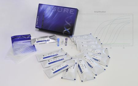
VIASURE Real Time PCR Detection Kits
Rhinovirus + Enterovirus

Description
VIASURE Rhinovirus + Enterovirus Real Time PCR Detection Kit is designed for the specific identification and differentiation of Rhinovirus and/or Enterovirus in respiratory samples from patients with signs and symptoms of respiratory infection.
This test is intended for use as an aid in the diagnosis of Rhinovirus and Enterovirus in combination with clinical and epidemiological risk factors.
RNA is extracted from specimens, amplified using RT-PCR and detected using fluorescent reporter dye probes specific for Rhinovirus and/or Enterovirus.
Specifications
Information
Human rhinoviruses (HRVs) and human enteroviruses (HEVs) are the most common cause of infections in people worldwide. They are members of the Enterovirus genus of the virus family Picornaviridae. They are small viruses (30nmin diameter) with an infectious single-stranded RNA genome of 7,000 to 7,500 nucleotides enclosed in an icosahedral capsid. HRVs include 153 currently known types divided into three species (A, B and C), while HEVs consist of 104 types belonging to four species (A, B, C and D). Traditionally, human enteroviruses are categorized into polioviruses and nonpolio enteroviruses (coxsackievirues, echoviruses, and numbered enteroviruses).
HRVs are the usual cause of common cold but are also frequently found in otitis media, sinusitis, bronchitis, pneumonia, and asthma exacerbations. Therefore, due to they are restricted to the respiratory tract the mode of transmission are mostly the via aerosols of respiratory droplets and from fomites (contaminated surfaces), including direct person-to-person contact. Currently there is no specific antiviral treatment for rhinovirus infection.
In contrast to HRVs, replication of HEVs is not restricted to the respiratory tract but also can take place in the small intestine and spread to various target organs. They are readily transmitted from person to person through an air and/or via a fecal-oral route, or even through contaminated objects. Most HEV infections are asymptomatic or manifest common cold-like symptoms. However, HEV infections can be more severe, causing poliomyelitis, meningitis, encephalitis, myocarditis, exanthema, acute hemorrhagic conjunctivitis, and severe generalized infections in newborns. Therefore, sample collection for HEVs diagnosis should be performed according to clinical manifestations. Cerebrospinal fluid (CSF), blood, respiratory samples and stool samples are commonly used.
Differential diagnosis of HRV and HEV infections is epidemiologically important. Specific identification of these viruses already has implications for the supportive management of patients and will become more significant when specific antiviral drugs become available. Nucleic acid amplification techniques have replaced the isolation of viruses in cell cultures as the method of choice for the detection of picornaviruses, partly due to the outstanding sensitivity, specificity, and rapidity of such techniques. The recently identified species C HRVs cannot be cultivated in standard cell lines but can be amplified by reverse transcription (RT)-PCR. Both HRVs and HEVs have conserved 5’ noncoding regions (NCRs) and a few nearly identical sequence motifs, allowing the design of universal primers for their amplification in RT-qPCR. RT-qPCR has been shown to be far more sensitive than cell culture for detection of these viruses.
